fbpx

Robotic Bypass Through a 4 cm Incision Without Opening the Thorax

Cardiovascular diseases are among the most common chronic diseases in our country. Bypass operations with robotic surgery for the treatment of cardiovascular diseases offer many advantages in terms of patient comfort and rapid recovery. Robotic bypass is possible by using 3 cuts in the armpit and a 4 cm incision on the rib cage without touching a scalpel to the heart or stopping it. Liv Hospital Cardiovascular Surgery Specialist Prof. Dr. Ahmet Ozkara: “We perform robotic bypass by using the most important vessel on the heart; thoracic aorta. Unlike conventional bypass surgeries, we complete the operation without opening the thorax and stopping the heart”. Highlighting that patients can return their normal life sooner with robotic bypass, Liv Hospital Cardiovascular Surgery Specialist Prof.
Dr. Ahmet Ozkara told that infection rates and bleeding is significantly decreased with this method, and said: “It is superior to conventional bypass surgery in terms of cosmetics as well. The patient can return to work within a few weeks. They reach normal physical conditioning.
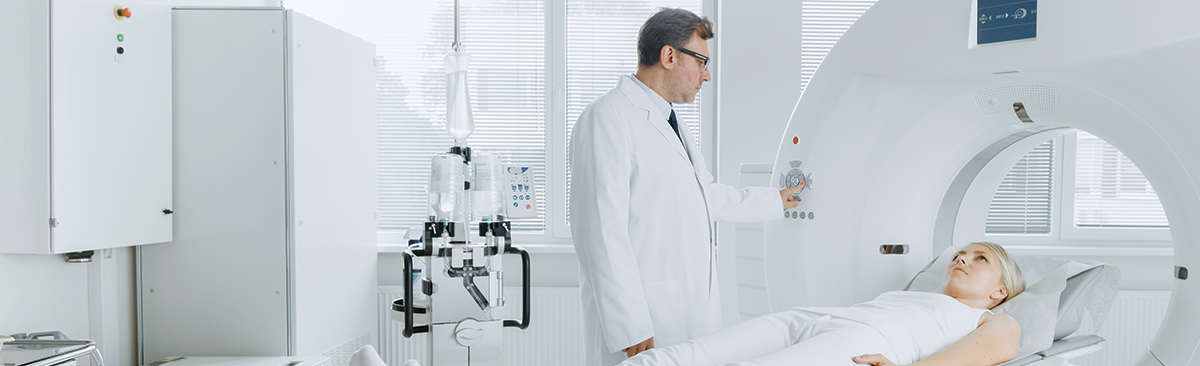
No scalpel is used.

With the technological developments of the last 15 years, the number of minimally invasive methods is increasing as an alternative to classical surgery methods in cardiac surgery. In the US and European countries, the number of robotic heart surgeries increased by 20 percent in recent years. And there is a similar trend in our country as well. In robotic cardiac surgery, the operation is performed without cutting the breastbone, the sternum. The robotic arm inserted through small cuts on right or left side of the rib cage. The 3-dimensional image obtained by the camera inserted through an incision provides a 8-10 times larger and clearer image. One surgeon attached the surgical instruments to the robotic arm, while another surgeon operates these instruments from a console with the camera display.

Comfortable for both patient and the surgeon

The instruments used in robotic surgery provide greater safety than the conventional or thoracoscopic surgery. The surgical instruments are operated by the finger movements of the surgeon at the console and these robotic arms are capable of movements that are difficult for wrist. There is no hand shake or fatigue, and intervention to anatomical regions with difficult access become easier. This provides comfort for both the surgeon and the patient.

Less pain, less infection

Coronary artery bypass surgery, mitral valve surgery, congenital heart surgery and arrhythmia surgery are performed by robotic surgery in eligible patients. Coronary artery bypass surgery is performed without stopping the heart or by stopping the heart (using a cardiopulmonary pump). Mitral valve operations are performed by stopping the heart with a special fluid. With robotic heart surgery, the recovery period is significantly shortened, and patients can usually return to work after 7-10 days. Bleeding, infection, pain and blood transfusion are less frequent. And it provides cosmetically superior results. 

Advantages of robotic surgery:

Better results:

  • Pain and some post-operative physical function disorders are less common, and better results are obtained in terms of patient satisfaction.
  • Less damage: The operation is performed through small incisions and with camera assistance; on the contrary to general belief, robotic surgery provides clearer and more detailed view than the open surgery, and this allows the operation to be completed with significantly less tissue trauma.
  • Faster recovery: The post-operative recovery is much more faster than open surgery. And this also translates into shorter hospital stay and faster return to normal life.
  • Less pain: As the surgery is performed through small cuts, patients feel less pain after the operation.
  • Less infection risk: Post-operative surgical site infection is less likely as the incisions are very small.
  • Better cosmetic outcome: Since the operation is performed with 1-1,5 cm cuts, better cosmetic outcomes are obtained in comparison to open surgery.
  • Better field of view: As the surgery is performed with the assistance of optical systems, more detailed and clear field of view is provided compared to open surgery.
  • Less blood loss: Blood loss is minimum thanks to less tissue damage.

Diplopia in Children

Normally, both eyes look at the same spot and vision axis are parallel to each other. The loss of this parallelism and two eyes looking at different directions is called diplopia (cross-eyed). The ability to track object-light with eyes and focusing the eyes on a target generally develops at third month. During this period, transient and small misalignments can be considered as normal. However, if there is a suspected misalignment after three months, then an ophthalmologist must visited. Liv Hospital Ulus Ophthalmologist Prof. Dr. Umit Beden explained diplopia.

Children and adults can have diplopia

Diplopia can be seen in both children and adults. Misalignment observed in childhood may not be related with another cause or can be accompanied by other conditions (cataract, eye tumors or neurological diseases) that disrupt vision. Therefore, eye examination is required immediately after diplopia is noticed. In childhood and infancy, diplopia can manifest with each eye focusing on different spots or closing one eye in dark and sunny environments or tilting the head to one side in daily life. And double vision is the most important symptom in adulthood.

Underdevelopment of the nasal bridge might give the appearance of diplopia in many babies. Therefore, it is wrong to assume that diplopia during infancy would recover in time. In case of such suspicion, the baby must be examined by an ophthalmologist as soon as possible. If there is diplopia indeed, it is unlikely to recover in time. Additionally, healthy visionary development of both eyes would be hindered in babies with diplopia and it is important to start treatment early to prevent this. Otherwise, lazy eye condition that can develop in one or both eyes will become permanent.

Normally, it is recommended that every healthy child should have their first eye examination between 6 months and 1 year. In addition to this, eye examination to be performed at the age of 3 is very important in terms of the treatment of any potential lazy eye condition. When diplopia is diagnosed, the goal of the treatment is to enable the healthy completion of the visionary development of the child. Therefore, eyeglass prescription, covering treatment and surgery can be planned. Instant treatment is not possible for childhood diplopia, a long-term follow up will be required. In case surgery is planned, it will not be an alternative to eyeglass prescription or covering treatment; eyeglass use and covering treatment may continue after the opeation.

360′ Overview of the Heart with Cardiac MRI

The heart is one of the most difficult areas to visualize due to its location inside the rib cage and behind the bone structures, and constant motion. Therefore, centers with state-of-the-art technology and experienced specialists are required for the detailed examination of heart-related functions and diseases. Stating that Cardiac MRI is an advanced imaging technique that enables a detailed examination of the heart without any interventional procedure, Liv Hospital Ulus Radiology Specialist Assoc. Prof. Dr. Ayse Gormeli explained the advantages of the method.

Cardiac MRI can be used for children

As no radiation is involved with Cardiac MRI, it can be safely used both for children and adults. And the drug (contrast agent) that can be used if necessary during the procedure is also significantly safer than the tomographic methods. This examination method provides the best possible calculation of the cardiac functions. The structures of the heart can be examined on every plane without any angle restriction. Pathologies in the heart can be clearly revealed.

For which conditions can it be used?

  • Cardiac MRI provides detailed information about the damage on the cardiac muscle in patients with vascular occlusion (vitality test). The extent of such damage is of crucial importance to determine whether the patient can benefit from a heart surgery.
  • In congenital heart diseases, Cardiac MRI offers significant contribution for pre-operative planning and risk classification by providing a detailed evaluation of the heart and large vessels. Additionally, it also allows for the acquisition of most reliable and accurate data in lifelong follow-ups after the surgery.
  • In cases of valvular insufficiency or obstruction, Cardiac MRI is used for the determination of the disease severity. The back flow rate of the blood through the valves can be calculated with a great accuracy.
  • It enables the discovery of underlying cause in cardiac arrhythmias.
  • In patients with suspected mass in the heart, it can be used to distinguish the mass and clot and to determine the malign tumors, if any.
  • Cardiac MRI also allows for the monitorization of the extent to which the heart is affected by evaluating the cardiac muscle in detail in case of diseases like mediterranean anemia (thalassemia), sarcoidosis, Anderson-Fabry disease, amiloidosis.

For which conditions it cannot be used?

Individuals with cardiac pacemaker or other non-MRI safe medical materials (prosthesis, cochlear implant etc.) are not eligible for MR imaging. In addition, it should not be used for claustrophobic individuals without proper medical assistance.

How does Cardiac MRI work?

A vascular access is opened on the arm before the examination. After the patient is taken into the MRI device, ECG leads are attached to the chest to see the cardiac motion. Breath holding exercises are performed as the patients are required to hold their breaths for short periods of time during the examination, and then the MRI acquisition is initiated. In cases where necessary, a drug called contrast agent can be administered from the vascular access. The examination is completed within 30-45 minutes on average.

Keep Your Intestines Happy and Cancer-Free with Mediterranean Diet

Colon cancer is one of the most common cancer both in the world and our country. Scientific studies have shown that Mediterranean diet, rich in vegetables, fruits, fish, olive oil, and nuts, significantly reduces cancer and vascular occlusion related deaths. Liv Hospital General Surgery Specialist Prof. Dr.
Hakan Yanar explained prevention, screening and treatment methods of colon cancer and the importance of nutrition.

What are the causes of colon cancer? Who are at risk?

More than 90% of colon cancers are caused by previously developed polyps. While the incidence of polyps is rare before the age 50, cancer rates increase thereafter due to higher polyp incidence. Nutritional habits that are rich in fat and low in fibers, colon cancer history in immediate family, inflammatory intestinal diseases like ulcerative colitis and Chron’s disease lead to increased colon cancer risk.

Is colon cancer dangerous?

Early diagnosis is very important in colon cancer, which is an asymptomatic and slow-progression cancer. This type of cancer is especially prevalent in people older than 50 and it can be treated with early diagnosis. Colon cancer is second or third most frequent cancer type in the world.

What are the symptoms of colon cancer?

  • Anemia
  • Constipation
  • Palpable mass
  • Rectal bleeding
  • Blood in the stool
  • Decreased stool diameter
  • Changed defecation routine
  • Inability to relief the urge to defecate
  • Abdominal pain for more than 15 days

How to diagnose early?

 Although occult blood in stool and immunohistochemical tests can be used for colorectal cancer screening, the most effective method is colonoscopy. The goal must be the removal of polyps, before they develop into cancerous tissues, during colonoscopy. Therefore, first colonoscopy screening must be performed at the age of 50 for people without colon cancer history in their family and, if the results are normal, it must be repeated every ten years. And for people with colon cancer in their family, first colonoscopy must be performed 10 years before the cancer onset age of the affected family member.

What is the treatment of colon cancer?

Standard treatment method is surgery. However, the removal of some healthy colon tissue and surrounding lymph nodes together with the tumors is of crucial importance. The surgical procedure affects long-term survival, relapse of the tumor or metastasis; therefore it must be performed by experienced surgeons. Today, these surgeries are performed with high success rates by using laparoscopy and robotic surgery methods, without any incision on the abdomen. Less post-operative pain, shorter hospital stay and very little scar around the surgical site are among the advantages of laparoscopic and robotic surgeries. In case of tumors close to the anus, colostomy (attaching the colon to the outer wall of the belly) can be prevented with chemotherapy and radiotherapy methods to be applied prior to the operation.

Follow the Recommendations

  • Have regular screening tests.
  • Consult your doctor in case of long-term constipation and rectal bleeding complaints
  • Do moderate intensity exercises for 4-5 days a week instead of an inactive life.
  • Apply a nutritional schedule that is rich in fibers, low in fats with balanced protein.
  • Avoid smoking and alcohol.
  • Maintain your ideal weight.
  • Scientific studies have shown that Mediterranean diet, rich in vegetables, fruits, fish, olive oil, and nuts, significantly reduces cancer and vascular occlusion related deaths.

Target-Oriented Irradiation In Prostate Cancer Treatment: 4-Dimensional Brachytherapy

Brachytherapy, one of the most important tools in radiation oncology, can be used for the treatment of prostate cancer. Brachytherapy addition can increase the efficiency of radiotherapy for patients who are not eligible for surgery in the organ-restricted stages of the prostate cancer. Similar to external irradiation, tumor is effectively destroyed with minimal side effects in brachytherapy. Liv Hospital Radiation Oncology Specialist Prof. Dr. Merdan Fayda explained 4-dimensional prostate brachytherapy.

Prostate brachytherapy allows irradiation of the cancerous tissue in the prostate from within, instead of external application. Therefore, very successful results can be obtained by targeting the tumor up close with high dose and without damaging the surrounding tissue.

4-Dimensional prostate brachytherapy

Liv Hospital Radiation Oncology Clinic offer real-time imaging guided high dose rapid prostate brachytherapy. HDR (high dose) prostate brachytherapy is performed with real-time transrectal ultrasonography for prostate cancer patients. Brachytherapy can be performed as an addition to external irradiation method IG-IMRT for suitable select patients.

What is the 4-dimensional high dose rapid prostate brachytherapy with real-time imaging guide?

The 4-dimensional prostate brachytherapy method is performed via plastic needles palced into prostate tissue under the guidance of transrectal ultrasonography. As the whole procedure is performed with real-time high-resolution ultrasonography guidance, it offers an extremely effective treatment planning and high accuracy application. The most effective treatment plan with least side effects is scheduled and delivered based on the real-time images of the prostate and other normal tissues. The procedure takes approximately 2 hours under general or spinal anesthesia and is completed after the removal of the needles. Then the patient is invited for external radiation or second prostate brachytherapy to be initiated one week later.

What are the advantages of prostate brachytherapy?

  • Total radiotherapy duration is shortened.
  • Treatment efficiency is increased.
  • The duration of hormone therapy can be shortened.
  • The damage of surrounding tissues and organs is reduced.
  • Less side effects.
  • Urinary incontinence/urinary tract stenosis incidences are significantly low.
  • It is usually performed in a single session and there is almost no risk of bleeding.

Who are eligible for prostate brachytherapy?

  • As an addition to the external treatment (IG-IMRT) of moderate and high-risk prostate cancer patients,
  • As focal radiotherapy in patients who previously received radiotherapy and had relapse,
  • As a single therapy for low-risk patients who are not eligible for selected operation.

What are the advantages of 4-dimensional high dose prostate brachytherapy over permanent seed brachytherapy?

  • The patient can return their normal daily life as there will be no radioactive source inside.
  • Real-time imaging guidance performed throughout the entire procedure increases the accuracy.
  • Since no radioactive source will be placed inside the patient, there is no displacement risk of such materials.

Protect the Rhythm of Your Heart

Although heart rhythm disorders are more common in the elderly, it is a heart condition that can occur at any age. This disease may have no vital risks, or lead to sudden death. Liv HOSPİTAL Cardiologist Prof. Dr. Enis Oguz explained what should be done to protect the rhythm of our heart.

What are the causes of cardiac arrhythmia?

Electrical stimulation is required for heart muscle to contract. The right atrium of the heart has cells that regularly outputs electrical stimuli. This electrical stimulus must spread all over the heart. The heart has lines consisted of sequentially located cells that the electricity flows through, as in an electrical grid. And the cardiac muscle responds to this electrical stimulus. Any abnormality in these structures results in the disruption of heart rhythm.
Arrhythmia exhibits itself in the form acceleration (tachycardia), irregular beat (premature beat) or decelaration (bradycardia).

What are the symptoms?

The most common symptom is the feeling of palpitation. Patients describe this feeling with statements like “My heart is like fluttering like a bird”, “As if my heart will dart out of my chest”, “My heart beats are visible from outside”, “I feel my heart beating in my mouth”. The palpitation usually starts suddenly and sometimes lasts for seconds, sometimes minutes and even hours, then stops. Other symptoms may include dizziness and fainting. Arrhythmia-related faints often indicate life-threatening types of arrhythmias.

Which age range is affected?

Arrhythmia can be seen in any age group. Arrhythmias are more common in older ages and are associated with other heart diseases. Taking all age groups into account, 2% of the general population and 10% of the population over 80 years of age is known to have atrial fibrillation (atrium-related arrhythmia of the heart). These numbers show the importance of cardiac arrhythmias for public health.

What tests are used for arrhythmia?
The most important method used for the diagnosis of arrhythmias is ECG, the recording of the electrical activity of the heart. However, in order for ECG to have diagnostic value, electrical activity must be recorded at the moment of arrhythmia. For example, if a patient suffers from an arrhythmia episode and goes to the nearest health institute while the tachycardia is ongoing to receive ECG, the type of the arrhythmia can be determined. However, some arrhythmias are short; therefore they cannot be recorded. For such cases, there are portable recording devices with the size of a cell phone, so patients can carry it with them. The purpose is to allow the patient to record their arrhythmia at the time it occurs.

3 years of cardiac rhythm recording

There are surgically placed subcutaneous recording devices, and they are used to record rare arrhythmias. This technique allows continuous arrhythmia record for 3 years. If these methods fail to identify the arrhythmia, then electrophysiological study, an interventional method, can be used. Some cases require diagnostic methods like echocardiography, cardiac MRI, coronary angiography. In addition, a special test is performed to identify the cause of fainting, called vasovagal syncope. In this disease, reflexive declaration of the heart rhythm and even temporary halt can occur. Differential diagnosis can be made with this simple method for many patients who are confused with epilepsy and who are under examination for diagnosis in neurology clinics.

Are patients with arrhythmia required to use specific diet?

Nutrition is important for patients with arrhythmia. Some substances posses stimulating properties for the electrical system of the heart. These may trigger arrhythmia. Effects of tea, coffee and smoking can be given as examples. In addition, many drug can also cause arrhythmia. Sometimes, heart and blood pressure drugs can cause bradycardia (deceleration). For example, drugs used for eye pressure can include substances that slows down the heart rate. Sometimes, a drug prescribed to treat an arrhythmia can cause another arrhythmia. General recommendations for cardiac health also apply for arrhythmia prevention. These include stress management, ideal body weight, regular exercise, nutrition without lard and trans fatty acid, reduced salt intake, quitting smoking, zero or reduced alcohol consumption.

What is Brachytherapy?

Brachytherapy is a type of radiotherapy where radioactive sources/radiation source is placed next to the tumor. It is being successfully used for the treatment of gynecological cancers (uterine, cervix, vagina), lung cancer, skin cancer. Today, brachytherapy can be applied as 3-dimensional with the use of imaging methods that are based on cross sectional anatomy like computed tomography and magnetic resonance imaging. As 3-dimensional brachytherapy is more frequently used, success rates increase and side effects decrease. Liv Hospital Radiation Oncology Specialist Prof. Dr. Merdan Fayda explained the brachytherapy method.

Brachytherapy has three fundamental application methods:

  1. Intracavitary brachytherapy: (Inside the cavities) performed by using applicators placed inside the natural cavities of the patient (uterine, cervix, bronchi etc).
  2. Interstitial brachytherapy: (Inside the tissue) performed by placing applicators inside the related tissues or by using radioactive sources delivered through a needle, as well as permanent placement of radioactive sources (permanent prostate uLDR brachytherapy).
  3. Contact brachytherapy: Radioactive source/radiation source is applied to external tissues such as kin or is applied at the time of removal of other organs during operation like in superficial intraoperative radiotherapy (IORT).

3 dimensional brachytherapy in gynecological tumors

In the treatment of gynecological tumors performed by physicians from different disciplines, brachytherapy has an important function and is applied by bringing radioactive sources near the area to be irradiated.

The most frequent area of use for brachytherapy is gynecological cancers. For uterus cancers (endometrium), cervical cancers and vagina cancers brachytherapy is applied after the operation, and in case of patients not fit for the operation, it is applied as single therapy method along with external irradiation.

Especially in the recent years, brachytherapy applications developed from 2-dimensional methods to 3-dimensional methods. Based on data acquired from computerized tomography and MR devices, 3-dimensional brachytheraphy applications contributes to the successful treatment of gynecological cancers and provides suitable protection for the surrounding tissues (bladder, rectum, sigmoid etc). This allows the treatment to be completed with less side effects.

Skin brachytherapy (with Leipzig applicator)

Brachytheraphy is successfully used for the treatment of early phase of skin squamous and basal cell cancers with a suitable depth and surface. Cosmetic damages arising from operation during the treatment of the tumors especially developing around the face are seen less with brachytherapy. Patients suffering from early stage skin cancer localized on the face may be treated with brachytheraphy if deemed eligible by the plastic surgeon.

3-dimensional brachytheraphy in lung (bronchi) cancers

In case of patients for whom external irradiation methods cannot be used in the radiotherapy of lung cancer, brachytheraphy can be performed through tubes to be placed inside the main airways with the assistance of pulmonology team.

In order to bring the radioactive source near the target area of the patient, certain assistant instruments such as applicators may be used. Although these applicators are mostly placed without any pain, sometimes anesthesia may be necessary.

A New Hope For Cardiac Patients: Dissolvable Stent

The dissolvable types of stent, medical devices used to keep cardiac vessels open, are becoming more common in recent years. Unlike metal stents, dissolvable stents do not remain inside the vessels for life. These stents start to dissolve after first 6 months and completely melts away in years, therefore the structure of cardiac vessels remains intact. Liv Hospital Cardiology Specialist Prof. Dr. Alp Burak Catakoglu explained the advantages dissolvable stent.

What are the symptoms of vascular occlusion or narrowness?

After the athersclerotis plaque, consisted of fat and calcification inside the coronary arteries, reaches a certain extent, the blood supply of the heart muscle becomes disrupted. Some typical symptoms may occur when the heart muscle is not supplied with enough blood and oxygen. The most frequently highlighted symptom is a punch-like sensation in the middle or left side of the thorax, or a pain in the form of compression or tightness in a wider area. In some patients, this may be felt in the jaw, back or left arm. These symptoms can be very mild or sometimes very significant. Many patients with cardiovascular diseases may not have these typical symptoms; sometimes, shortness of breath during effort or premature fatigue can be the first signs of this disease. It may also progress quietly and stealthily in some patients, and a heart attack may be the first time that the disease reveals itself. Severe chest tightness can be accompanied by shortness of breath, cold sweat and fainting. In case of such condition, it is absolutely necessary to apply to the nearest health institution as quickly as possible.

What happens if vascular occlusion is not treated?

According to World Health Organization (WHO) data, cardiovascular diseases are the most lethal diseases in the world. As the coronary vessels start narrowing, it may cause a heart attack in the future or may result in sudden death. People with cardiovascular disease, diabetes, hypertension, or high cholesterol or smoking in their parents or siblings may experience cardiovascular disease at an earlier age. Patients with increased cardiovascular risk in this group should be checked more carefully and frequently. Studies suggest that diabetic patients are at a four times higher risk of heart attack. In order to reduce the risk, it is very important to assume diabetic patients as cardiovascular patients from the beginning and regularly review their treatment with a cardiologist. Early intervention can save patients who have a heart attack, while it may result in the death of some patients or they may suffer from severe damage to cardiac tissue and have to struggle with heart failure. Therefore, the main goal is to diagnose cardiovascular disease at an early stage, before a heart attack, and to take necessary precautions.

What diseases does vascular occlusion cause?

Vascular occlusion can lead to heart attacks, cardiac failure, rhythm irregularities and death. Vascular occlusion and stenosis can be diagnosed at an early stage with the present technology. Depending on the risk profile of the patient, cardiologists can start the treatment at an early stage and determine the severity of the disease by use of many advanced test methods.

In which situations can the dissolvable stent be used?

The use of dissolvable stents can be considered for almost all patients who are eligible for metal or drug-coated metal stents. And it has additional advantages for some patients. It is recommended for patients who are to receive stents for the first time. Makes it easier to apply future treatment to the same area. Young patients benefit more. It should be preferred for patients who are likely to undergo the procedure again in the future. Recurrent occlusion and repeat treatment should be taken into consideration in patients with risk factors. Therefore, metal stents may require second metal stent placement (sometimes even for the third time). In particular, diabetes, hypertension, smoking, high cholesterol and cardiac disease history in the family increases this possibility. Instead of standard angiography, coronary tomography can be used for the follow-up of the stented area, therefore patient comfort and safety is increased. Application of metal stent to patients with metal allergy increases the likelihood of repeated occlusion. It can be applied for thin vessels and long-term diseases area, which may not be eligible for bypass surgery.

What are the advantages of dissolvable stent?

Coronary vessels’ stent need is for short-term. There is no advantage in leaving the stent inside the vessel after its task is completed.
After the stent is dissolved, the vascular structure remains intact with normal functions. It does not hinder future treatments.
Re-stenting the same area or bypass operation may pose a few challenges.
Blood thinner drug use is shortened.
It is impossible to examine an area with metal stent by using tomography. However, vessels with dissolvable stent can be visualized with coronary tomography. It increases quality of life.

Radiotherapy Treatment In 8 Questions

Although the incidence of cancer continues to increase, new hopes emerge with new treatment methods and technological development. In recent years, radiotherapy application with advanced technological devices is considered as one of the most effective methods in cancer treatment. Liv Hospital Radiation Oncology Specialist Prof. Dr. Merdan Fayda explained radiotherapy treatment.

What is radiotherapy (radiation therapy)?

Radiotherapy is a treatment method with ionized radiation. In fact, we are not stranger to the energy transmission in the air. Many device, such as mobile phones, television antennas, satellite positioning systems (GPS) and radios, work according to the principle of energy transmit in the air. The medical treatment we apply with beams is called “radiotherapy” or “radiation therapy”. The beams used for radiotherapy are ionizing; they have the power to damage the genetic material, DNA. Applied radiation damages the tumor DNA as well as the normal tissues. Tumor cells cannot survive the damage caused by radiotherapy, however, normal tissues can repair such damage. Therefore, radiation is being successfully used for therapeutic purposes since its discovery in 1890s.

What is felt during radiotherapy (radiation therapy)?

The entry and exit of the low-energy X-rays used in tomography is not perceived by the patients. High-energy beams used in radiation therapy are also not perceived. Therefore, radiotherapy patients do not feel much during the application.

Are the relatives of patients going through daily radiotherapy affected by the radiation?

The radiation applied during radiotherapy is generally performed by external radiation devices which rotate around patients while they lay without moving. As these beams enter and exit the body at the speed of light, they do not accumulate and therefore there is no risk of exposure for patients’ relatives. In brachytherapy (close radiotherapy), another form of radiotherapy, apparatus that deliver radioactive sources inside the cavernous organs or tissues (e.g. Uterus) of the patients are used. After these apparatus are placed inside the patient, radioactive source travels to the region to be irradiated and irradiation takes place. Then, the radioactive source is released from the apparatus. The patients are sent back home after the apparatus are removed. As there is no radiation accumulation after brachytherapy, patients’ relatives are at no risk of radiation exposure.

What are pre-radiotherapy preparations?

Radiotherapy can be performed at very different stages. Sometimes, it may necessary to complete certain tests before the treatment. Before starting, the reason for radiotherapy need is explained to the patient by a radiation oncology specialist. Most of the radiotherapy applications will cover the entire organ or a part of it as in surgical practices, or the lymph nodes where lymph fluids is discharged. Therefore, different preparations may be required for different regions. For example, preventive oral care is recommended for patients who are preparing for head-neck radiotherapy. Patients preparing for lower abdominal radiotherapy may be adjusted for fullness or emptiness of the bladder and rectum. Blood count may be required before starting the treatment since blood count values will be important during radiotherapy.

What determines the radiotherapy duration?

Radiotherapy planning usually starts with computed tomography. The radiation oncologist, who will decide the area to be irradiated, makes the necessary drawings on computed tomography. Arrangement of the areas required for irradiation of the  targets and how to irradiate them are decided together with the medical radiophysics specialist. This is how the time our patient will spend each day in the treatment device and for how long it must be performed is determined. The patient’s history of surgery, the condition of the disease, details of the pathology report and other accompanying diseases of the patient are important for the determination of treatment duration.

What are things to watch out during radiotherapy?

Radiotherapy applications are performed under the supervision and control of the radiation oncology specialist. The recommendations of the physician should be followed and controls should be performed during radiotherapy. During radiation therapy, the physician should be informed before using any medication, especially in cases with newly developed complaints such as fever, difficulty in swallowing.

How should be the nutrition during radiotherapy?

Nutrition during radiotherapy may vary depending on the irradiated area. For example, in the radiotherapy involving the head and neck and esophagus, a diet consisting of soft foods is recommended, whereas in the radiotherapy for lower abdomen, a diet that will not cause diarrhea is preferred. As some cancers like breast cancer are sensitive to hormones, physician may recommend against weight gain. It will be helpful to ask the physician about nutritional considerations during radiotherapy.

Is social life affected by radiotherapy?

During radiotherapy, social life is not affected as much as chemotherapy. However, it is necessary to consult a doctor. For example, the doctor should be consulted to learn whether meeting with people other than the family will harm the patient. Sharing some information about the disease with family and relatives may be helpful for the patient to pull through radiotherapy and other treatments.

Robotic Lung Cancer Treatment In 5 Questions

What is the difference between robotic surgery and open and other close surgeries?

In surgical lung diseases and lung cancer, robotic surgery or robot-assisted surgery provides more accuracy, flexibility, control and safety for surgeons in complex procedures compared against conventional methods. As the lymph node removal in lung cancers is performed more successfully then video-assisted surgeries, robotic methods provide oncological advantages. Robotic surgery is a type of closed surgery called minimally invasive, which are performed through small incisions. In thoracic surgery, the superiority of video-assisted of thorascopic closed operations over open surgeries has been proved. After the da Vinci Robot was approved by FDA in the US back in 200, it rapidly became a common practice in American and European hospitals for the treatment of many diseases in various fields. Robotic surgery involves a camera arm, and mechanic arms where surgical tools attached. The surgeon sits at a computer console near the operating table and performs the operation by controlling these robotic arms. The console provides the surgeon with a high-resolution, magnified and 3D image of the surgical site. Surgeon directs the assisting operation crew from the console table.

What are the advantages?

In comparison to conventional methods, surgeons using the robotic system have higher accuracy, flexibility, control and better view of the surgical site during the operation. The tools used with the robotic system provides the flexibility of a wrist inside the thorax. With the robotic surgery systems, surgeons can perform sensitive and complex procedures that could be difficult or impossible otherwise.
Advantages of minimally invasive robotic surgery are: Less
Complications (e.g. Wound site infection), less pain, less blood loss, faster recovery, smaller and less noticeable incision scars.

Are there any risks?

In robotic surgery, similar complications in conventional methods are less prevalent. Robot equipment allows for minimal damage to the surrounding tissues within the thoracic cavity and reduces the complication rate.

Is every patient eligible?

Robotic surgery may not be an option for all patients. The surgeon explains the patient whether they are eligible for robotic surgery, the benefits and the risks by comparing it with other methods (open surgery and video-assisted surgery).

For which diseases it should be preferred?

Robotic surgery can be reliably and safely used for many diseases in thoracic surgery. These include early stage “lung cancers”, tumors and cysts in the area between two lungs within the thoracic cavity called “mediasten”, “diaphragm diseases”, “thymoma”, “Myastenia Gravis” (thymectomy). As the lymph node removal in lung cancers is performed more successfully then video-assisted surgeries, robotic methods provide oncological advantages.